2015 Hot Selling Laptop Ultrasound
USD $1 - $5,000 /Unit
Min.Order:1 Unit
Quick Details View All >
Guangzhou Joyful Technology Co., Ltd.
Product Details
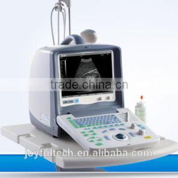
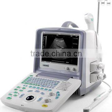
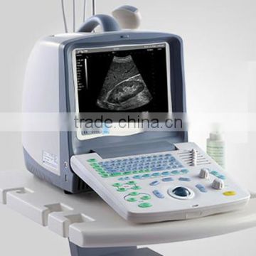
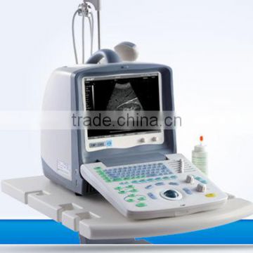
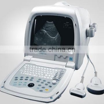
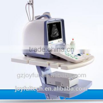
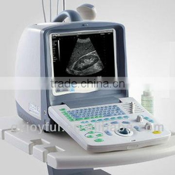
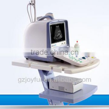
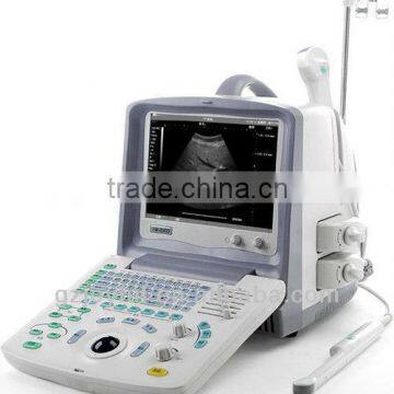
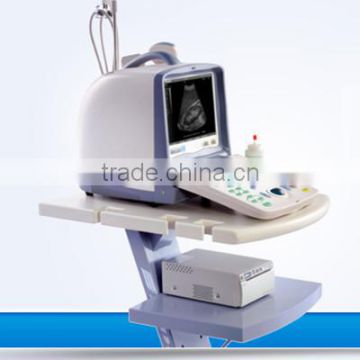
2015 Hot Selling Laptop Ultrasound
Overview
Imaging mode: B, B+B, 4B, B+M, M
Gray scales: 256
Display: 10 inches non-interlaced SVGA
Probe frequency: 2 ~10MHz
Probe connector: 2 standard
Beam-forming: Digital Beam-forming
Dynamic Receiving Aperture
Dynamic Frequency Scanning
Dynamic Receive-focusing
Dynamic Filtering
Scanning angle: From 30 to 120 degree (depending on probes)
Scanning depth (mm): From 40 to 250 (depending on probes)
Detail Description
1 Imaging Processing:
Pre-processing:
Pre-settings, Dynamic range, Edge enhancement, Frame correlation
Line correlation, 6-segment TGC adjustment, 4-focus adjustment
2 Post-processing:
Gray transform, Gamma correctin, Rejection, Left-right reverse
Up-down reverse, Polarity reverse
Min. 11 levels zoom, Partial zoom, Real-time histogram
3 Functions:
Cine loop: 365 frames bidirectional cine-loop
Storage media: Built-in flash, external pen drive
Storage: Min. 50 frames permanent storage and 16-frame snapshot
Body mark: 64 types
Probe auto-detection
10-level acoustic power output adjustment
4 Measurement & Calculation:
B-mode: Distance, circumference/perimeter, area, volume, angle, hip joint, stenosis ratio
M-mode: Distance, time, velocity, EF slope, heart rate
Software packages: Abdomen, gynecology, obstetrics, urology, small parts, cardiology
5 Standard Configuration:
Main Unit
10" Non-interlaced SVGA
365 Frames Bi-directional Cine Loop
Min. 1600 Frames Permanent Storage
16 Frames Snapshot
Body Mark: 64 Types
Tissue Harmonic Imaging(THI)
Tissue Specific Imaging(TSI)
Professional Software Packages
2 Transducer Connectors
2 USB Ports
6 Optional:

What is the theory?
Ultrasound images (sonograms) are made by sending a pulse of ultrasound into tissue using an ultrasound transducer (probe). The sound reflects (echoes) from parts of the tissue; these echoes are recorded and displayed as an image to the operator.
How about Ultrasound images?
Compared to other prominent methods of medical imaging, ultrasonography has several advantages. It provides images in real-time (rather than after an acquisition or processing delay), it is portable and can be brought to a sick patient's bedside, it is substantially lower in cost, and it does not use harmful ionizing radiation.
Contact Supplier

You May Like
New Products


Popular Searches
Recommended Products
Find Similar Products By Category


 Facebook
 X
X
 Pinterest
 Linkedln